
Arctic Fox in its full fluffy white winter glory. All best wishes for a happy, healthy, and successful new year! And keep snurfling. Image credit: Eric Kilby via Flickr.
Arctic Fox in its full fluffy white winter glory. All best wishes for a happy, healthy, and successful new year! And keep snurfling. Image credit: Eric Kilby via Flickr.

This rather bedraggled looking mess is an Arctic Fox (Bering Island subspecies) beginning to shed the dark summer coat. Image credit: A. Shienok / United Nations Development Programme in Europe and CIS via Flickr.

Image credit: Yale Peabody Museum / Curious Sengi.
While drifting through the Invertebrate Paleontology collections one day, I found a tray of lovely trilobites, many of them surrounded by a golden yellow halo.

Image credit: Yale Peabody Museum / Curious Sengi.
Though I do not know enough about these specimens to say what caused these halos, it is likely some kind of iron oxide stain produced by a chemical reaction between the surrounding rock material and the organic stuff oozing out of the trilobite during the fossilization process. In any case, this quirk of preservation gave these trilobites a rather ethereal glow. . . . .
Let’s have some fun with that!

Trilobites are the ultimate Trinity. They get their name from the three lobes that divide up the main body: a central axial lobe with two pleural lobes on either side. Apologies to The Nativity of the Lower Church at Assisi by Giotto (c. 1306 – 1311). See the original fresco here.

Isn’t that better? No judgmental babies here. Just a glorious cephalon. Apologies to La Vierge au lys by Bouguereau (1899). See the original painting here.
But sometimes Nature shows you something simple and evocative. No cheap tricks and ersatz Photoshopping necessary. What do you see here? A tender “mother and child” pose? A random assemblage of bodies? One trilobite headbutting another?

Image credit: Yale Peabody Museum / Curious Sengi.
No matter how you view this image and how ever you will be celebrating this time of year, all best wishes from the Curious Sengi! 

Preserved specimen of the deep sea hydroid, Branchiocerianthus imperator, is kept afloat in its jar by an air-filled glass float. Image credit: Natural History Museum of Denmark / Curious Sengi.
It was an object which was calculated to raise enthusiasm in a naturalist. A large disc surmounted a long stalk which evidently fixed the animal on the sea-bottom. A circle of numerous graceful tentacles hang down from the margin of the disc. . . . and the prevailing colour transparent scarlet (Miyajima 1900).
What Miyajima was describing is a specimen of Branchiocerianthus imperator, a solitary hydroid brought up by a long-line from a depth of over 450 meters (~1500 ft) off the coast of Japan. Hydroids are cnidarians, a group of aquatic invertebrates that use specialized stinging cells to capture prey and include more familiar creatures such as jellyfish, sea anemones, and corals. Most hydroids are small and colonial, but B. imperator is a majestic loner looming nearly a meter (~3 ft) over the deep sea plains of soft sand and mud (Miyajima 1900; Omori & Vervoort 1986).

Example of a more typical solitary hydroid. This individual is likely no more than a couple of centimeters in diameter. Image credit: Dawn Clerkson via Scubaverse.com.

B. imperator as spotted by the Japanese submersible Shinkai 2000. These wonderfully strange animals consist of the hydrocaulus “stalk” and the hydranth “flower”, which bear the stinging tentacles used to capture food particles and small prey items. Miyajima also noticed that unlike many other cnidarians that have radial symmetry, B. imperator possesses bilateral symmetry (Miyajima 1900). Image credit: Oneclickwonders blog.
Delighted by the delicate reds and pinks of the animal: “It was agreed on all sides that it was a New Year’s gift from Otohime and that it should be known in Japanese as Otohime no Hangasa.” Though Miyajima was writing for a Japanese academic journal, he included a footnote to explain the origins of the moniker: “‘Otohime’ is a beautiful goddess who is supposed to have her palaces at the bottom of the sea. ‘Hanagasa’ is the flower-sun-shade or ornamental parasol. Thus Otohime no Hangasa means ‘the ornamental parasol of Otohime.’” Eager to preserve the color, Miyajima and colleagues placed the hydroid in a formalin solution, only to be disappointed as the tissues slowly bleached white (Miyajima 1900).

Examples of other hydroids illustrated in artistic exuberance by zoologist Ernst Haeckel. B. imperator is not represented here, but its closest relatives are the tall slender hydroids flanking either side of the plate. Omori and Vervoort (1986) described the living B. imperator as giving “. . . .the impression of the flower of a daffodil on its long stalk.” Image credit: Ernst Haeckel, “Kunstformen der Natur” (1900) via BioLib.
Despite its discovery in 1875 during the famous HMS Challenger expedition, very little is known about B. imperator. The bleached tissues and rather bedraggled look of preserved specimens have yielded important anatomical and phylogenetic data, but little else. This hydroid has been found in West Pacific waters, especially off the coast of Japan, at depths of 50 to 5307 meters (164 – 1739 ft) which make it particularly difficult to study. But brief observations of the live animal by scientists in submersibles indicate some interesting facets to the life of the largest known solitary hydroid. A juvenile myctophid fish, approximately 15 – 20 mm long, was captured by the trailing tentacles and after a 90 second struggle, was subdued by the sessile predator. In addition, tiny red shrimp were observed living symbiotically at the base of the tentacles, but their relationship with the hydroid remains a mystery (Omori & Vervoort 1986).
Buoyed by a glass float (perhaps a coincidental nod to Japanese glass fishing floats sometimes found by beachcombers) this particular specimen of B. imperator shows the whole outstretched length of the animal. Called “The Emperor Polyp” on the museum label, this item was purchased in 1914 from a dealer in Japan by Danish marine biologist Theodor Mortensen. I do not know how this individual was preserved, but some of that original rosy glow remains in the hydrocaulus, or “stalk”, of this specimen. Miyajima would undoubtedly smile at this presentation of the goddess Otohime’s flower parasol.

Image credit: Natural History Museum of Denmark / Curious Sengi.
Miyajima, M. 1900. “On a Specimen of a Gigantic Hydroid, Branchiocerianthus imperator Allman, found in the Sagami Sea.” The Journal of the College of Science, Imperial University of Tokyo 13: 235 – 262.
Omori, M. & Vervoort, W. 1986. “Observations on a Living Specimen of the Giant Hydroid Branchiocerianthus imperator.” Zoologische Mededelingen 60 (16): 257 – 261.

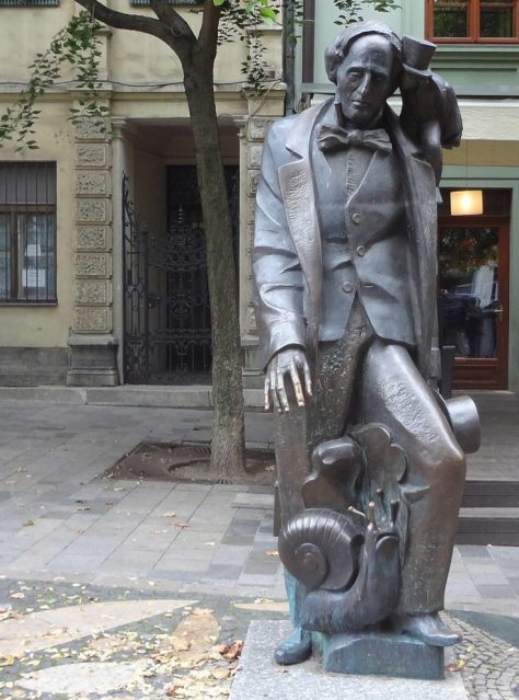
Danish author Hans Christian Andersen, who is most famous for writing beloved fairy tales such as “The Little Mermaid”, “Thumbelina”, “The Snow Queen”, “The Ugly Duckling”, and “The Emperor’s New Clothes”. Image credit: Thora Hallager via Wikipedia.
The Curious Sengi recently spent a few days in Copenhagen, snurfling about that wonderful old city. Going to visit the iconic statue of The Little Mermaid (Den lille Havfrue) was nowhere near the top of my list of things to do, but since I was already walking the ramparts of the star-shaped citadel of Kastellet, I dropped down to the harbor below to take a look. For a brief moment, I saw her: alone, looking sadly across the murmuring waves. But even the wet chill of mid-autumn did nothing to deter tour buses from roaring up the drive and disgorging scores of tourists. They gawk. They pose with big smiles and snap selfies. For a few, maybe this is the fulfillment of a dream. For others, it is just another check mark closer on the itinerary to lunch. But standing back and watching the rowdy proceedings, you cannot help but feel a bit pained. She’s such a poor little slip of a thing, caught in the moment of her greatest despair.

These daily disturbances are the least of the insults The Little Mermaid statue has endured. She has been variously decapitated, dismembered, blown up, and drenched in paint. As an easily accessible and visible tourist attraction, the statue has become the focus of protest statements and simple vandalism (Wikipedia 2016). Image credit: Curious Sengi.
Though there is a growing awareness that the fairy tales put on the big screen by Disney are intensely sugar-coated variants of the original stories that inspired them, I had to wonder how many of those tourists really knew “The Little Mermaid”, written by Danish author Hans Christian Andersen (1805 – 1875). I certainly had to refresh my memory. The story is bleak. Let’s just say that after being abandoned by the prince for whom she had sacrificed so much, the only happiness the Little Mermaid will ever experience is the release of death and the promise of an immortal soul. . . . . or disintegration into sea foam. I assume there is no noteworthy distinction there. Andersen’s story is complex, haunting, and even ambiguous. Then again, so was Andersen himself.
A vignette of Andersen’s life can be found in a small collection of shells at the Natural History Museum of Denmark in Copenhagen. He began collecting local land snail shells because of Jonas Collin, the son of his close friend, Edvard. Jonas had taken up zoology and enthusiastically collected snails, an endeavor that filled their lodgings with the stink of the boiled creatures and preservative spirits when Andersen and Jonas traveled together in 1861.

Shells of land snails collected by Andersen during his travels throughout Denmark. The Natural History Museum acquired this collection in 1905. Image credit: Natural History Museum of Denmark / Curious Sengi.
During this journey, the much younger Jonas proved to be “harsh, insulting and assertive”; the constant arguments and ungrateful attitude on Collin’s part was shattering to Andersen, who confided his tearful feelings to his diary. But within this tumultuous relationship between the imperious young naturalist and the older doting author, there were moments of happiness. At the end of their travels together, the pair parted amicably and Andersen would continue poking around gardens with newly acquired interest to find more specimens to send to his friend. Andersen included this note in a letter to Jonas’ mother:
Yesterday I found a beautifully coloured snail. I thought immediately of Jonas and took it up to my room, then went to lunch; but when I went back upstairs, the snail was nowhere to be found. I searched and searched, then found the beast had crept up the back of the table. I put it in my soap dish and put a lid over it. Last night, I wanted to make my specimen, boil the snail, remove the body and keep the shell for my scientific friend, but when I came to retrieve my victim, it had disappeared again, and this time it was lost for all time. The maid had cleaned up and thrown the snail out the window, but Jonas will see that I was thinking of him, and that will appeal to him (Andersen to Henriette Collin 5 June 1862; quoted from Strager 2014).
Upon hearing the news, Jonas responded rather like a pedantic brat:
That you collect snails for me is very touching, but the fact that you let them run away again, in my opinion, balances out the praiseworthy. I would very much appreciate snails from Basnaes. You can keep them alive in a dry box with small holes in the lid. They survive for years without food by sleeping (quoted from Strager 2014).

Image credit: Natural History Museum of Denmark / Curious Sengi.
Though Andersen’s snail collecting eventually slowed to a halt (Strager 2014), he was mindful of pleasing Jonas, almost to an obsequious degree. Sometimes the young man expressed gratitude, but it can be puzzling why Andersen was so invested in this unbalanced relationship.
One possible way of understanding Andersen’s relationship with Jonas is to acknowledge the intense, and ultimately unreciprocated, feelings that Andersen had for Jonas’ father, Edvard. There can be a long discussion about whether or not Edvard Collin was once seen as a lover (Bom & Aarenstrup 2015), but it is clear enough that Andersen put both men and women at the center of his romantic and erotic attentions. In his private writings, Andersen expresses his deep yearnings, all of which seemed to end in rejection and celibacy (Bech 1998; Bom & Aarenstrup 2015).

Andersen with Jonas Collin, around the time of their snail collecting venture. Image credit: Bordeux Barberon via Wikimedia Commons.
I wonder if during his rambles with Jonas, Andersen learned that snails are hermaphrodites, capable of reproducing as both males and females, sometimes simultaneously. Andersen seems to have seen himself as androgynous (Bom & Aarenstrup 2015). Natural history would hint at a sympathetic connection between Andersen and snails — another unassuming, ugly duckling-like character that Andersen identified with. And yet he cast the snail as an egocentric curmudgeon in the story “The Snail and the Rosebush” as a jab against certain philosophers in his acquaintance (Strager 2014). No soft spot for snails there and, thus, our natural history fairy tale falls apart.
Like “The Little Mermaid”, the story of Andersen’s life was filled with much more pain and complexity than is perhaps understood. His collection of snail shells in the Natural History Museum of Denmark is but a small glimpse of that life.
Statue in Bratislava, Slovakia commemorating Andersen and some of his famous fairy tale characters, including the grumpy snail. Image credit: weepingredorger.
Read Andersen’s short story: “The Snail and the Rosebush”
Bech, Henning. 1998. “A Dung Beetle in Distress.” Journal of Homosexuality 35 (3-4): 139 – 161.
Bom, Anne Klara & Anya Aarenstrup. “Homosexuality.” H.C. Andersen Centret: FAQs. Syddansk University, 11 August 2015. Accessed 27 November 2016.
Strager, Hanne. 2014. Precious Things: The greatest treasures of the museum. Tam McTurk, translator. Gylling: Narayna Press.
Wikipedia contributors. “The Little Mermaid (statue).” Wikipedia, The Free Encyclopedia. Wikipedia, The Free Encyclopedia, 24 November 2016. Accessed 27 November 2016.

Illustration of two European mustelids, the Belette (Mustela nivalis) and L’Hermine (Mustela erminea), from Volume 7 of Illustrations de Histoire naturelle générale et particulière avec la description du Roy (1758). James Madison consulted this work when describing his own locally-caught weasel. Image source: Wikimedia Commons.
Just a few years before authoring the U.S. Constitution’s Bill of Rights, James Madison (1751 – 1836) was sitting at home in his Virginia estate, dissecting a weasel and writing up his detailed results in a letter to Thomas Jefferson (1743 – 1826).
This letter — dated June 19th, 1786 — is a remarkable one, the sort of enlightened discourse one imagines of such great minds. It begins with a discussion about the nature of poverty in Europe (described by Jefferson in a previous letter) and the United States, as well as the presumptive role the mode of government had in shaping the existence of the poorer classes. A little report on the weather and the crops, then Madison expresses “. . . a little itch to gain a smattering in chymistry. Will you be kind eno’ to pick up some good elementary treatise for me. . .[?]” There is a brief paragraph on pushing through a state legislative bill for road repair and maintenance. Then come the weasels.

James Madison, statesman of Virginia, co-author of the “Federalist Papers”, architect of the Constitution and Bill of Rights, 4th President of the United States, dissector of weasels. Image credit: Gilbert Stuart: The Complete Works.
Madison explains that the body of a female weasel (Mustela frenata) came into his possession and then continues to fill up over two pages (in this four page letter) with detailed descriptions of the animal. Here is a taste:
Its colour corresponded with the description given by D’Aubenton of the Belette & Roselet or Hermine in its summer dress, excepting only that the belly &c. which in the European animal was white, was in ours of a lightish yellow, save only the part under the lower jaws which was white for about ½ an inch back from the under lip. The little brown spots near the corners of the mouth mentioned by D’Aubenton were peninsular. The tail was of the color of the back &c. all but the end which was black. The ears were extremely thin, had a fold or duplication on the lower part of the conque about 2 lines deep, and at the margin all around were covered with a very fine short hair or fur of the colour nearly of the back. The rest of the ear was in a manner naked, and of a lightish color.
Madison just keeps on going. . . . and these were not just superficial observations. The man measured weasel kidneys in three dimensions and counted the number of ridges in the palate. Whether to wife Dolley’s consternation or approval we may never know, but Madison was digging into the anatomy of small mammals with the same sort of intensity with which he approached national constitutions.

An example of the beast in question: the Long-Tailed Weasel (Mustela frenata). Weasels are fierce little carnivores, occasionally taking down prey many times their own size. Image credit: Evan Jenkins via Flickr.
There is little doubt that Madison was emulating his correspondent and role model Jefferson by drawing up charts comparing a variety of anatomical measurements between his weasel and the European “Belette” and “Hermine.” These tables of measurements reflect those published by Jefferson the previous year in his Notes on the State of Virginia (1785). What Madison was doing was comparing the anatomy of his American weasel with European species as described by the natural history authority, Louis Jean Marie Daubenton (1716 – 1800), for the purpose of building up evidence against Old World ideas about biology. Madison’s specific conclusion from his dissection of the weasel:
The result of the comparison seems to be that notwithstanding the blackness of the end of the tail & whiteness of the feet, which are regarded as characteristics of the Hermine contradistinguishing it from the belette, our weasel cannot be of the former species, and is nothing more than a variety of the latter. This conclusion is the stronger, as the manners of our weasel correspond more nearly with those of the Belette, than with those of the Hermine. And if it be a just conclusion, it may possibly make one exception to Buffon’s position that no animal is common to the two continents that cannot bear the climate where they join; as it certainly contradicts his assertion that of the animals common to the two continents, those of the new are in every instance smaller than those of the old.
In this last statement, Madison is referring to prominent French naturalists such as Daubenton and the Comte de Buffon (1707 – 1788) who claimed that America was only capable of producing puny counterparts to European species. This notion of degeneracy was summarized by Jefferson in Notes on the State of Virginia:
The opinion advanced by the Count de Buffon is 1. That the animals common to both the old and new world, are smaller in the latter. 2. That those peculiar to the new are on a smaller scale. 3. That those which have been domesticated in both, have degenerated in America: and 4. That on the whole it exhibits fewer species. And the reason he thinks is, that the heats of America are less. . . . that heat is friendly, and moisture adverse to the production and development of large quadrupeds.
It did not take much imagination to figure out that degeneracy applied not just to animals, but to people as well. This was a humiliating and detrimental statement against a newly-birthed nation striving to assert its independent identity and ability to thrive in the face of global skepticism. It was also blatantly contradictory to what Americans observed daily on their farms, in the forests, and in the ranks of their own fellow citizens. Men like Madison and Jefferson — as well as Benjamin Franklin, John Adams, and Alexander Hamilton — were infuriated by such pretentious Old World assumptions about the New World (Dugatkin 2009), assumptions that were not based on records of observed facts, but upon some kind of arbitrary, feel-good narrative. No sophisticated argument was necessary to simply show that North American animals are not categorically diminutive compared to European ones. But it was still important to make sure those data were carefully collected and circulated. Eventually, the idea of degeneracy lost steam and died out.

Madison’s letter to Jefferson included this rather spectacular table of measurements comparing his weasel (left column) with European species (middle and right columns). As Mythbuster Adam Savage said: “Remember kids, the only difference between screwing around and science is writing it down.” Well, honestly, I cannot imagine Madison screwing around. Image credit: Library of Congress.
I wonder if it should be remarkable to us in this modern age, to learn that these men who led the American Revolution and the early days of the Republic — many of whom would serve as President of the United States — engaged in what we would recognize as science. They were not necessarily scientists, though they thought scientifically. Science also had a broader meaning in the 18th century, a meaning that stood at the foundation of the Enlightenment ideal. Science was the application of empirical, experimental studies for the betterment of the human condition. Science was the triumph of reason and Nature’s laws over fanaticism.
Take, for example, wealthy farmers such as Jefferson, Madison, George Washington, and John Adams. They were all involved in agricultural research of some sort, keeping extensive multi-year records in an effort to develop better compost mixes, meteorological predictions, plant cultivars best suited for a given climate, and methods of crop rotation (Engle 2002; Druckenbrod et al. 2003; “George Washington and Agriculture”). Of course, increasing crop yields did contribute directly to personal gains in wealth, but it was also about making gains in national wealth. As gentlemen farmers, these men felt an obligation to be the experimenters because they were in a better position to absorb the costs of failure impossible for smaller subsistence farmers (“George Washington and Agriculture”). What they learned, they shared in the interest of making the new United States a profitable and self-sustaining continent. In turn, this made the largely isolationist policy of the Early Republic possible during those vulnerable, fledgling years.
Science had a real impact on the fate of the United States. Jefferson, Madison, Washington, Adams, and their compatriots understood this.
Science continues to have a real impact on the fate of the United States and the world, but I am much less confident that our elected leadership understands this.
For all the things we may find distasteful, hypocritical, or abhorrent about the 18th century world that produced the Age of Enlightenment, I think we still must admire the dedication to the painstaking business of improving the state of humankind through reason, sympathy, and a better understanding of Nature. Certainly, science has changed a lot since the days of Madison dissecting a weasel at home in Montpelier. A lot of research now requires specialized facilities and training only available through higher level university education. There are increasingly more specialized niche fields with their own language and communities, each one producing more published literature than can possibly be consumed or understood. But this is no excuse to reject science. This is no excuse to ignore the thousands upon thousands of scientists reaching out to the public and shouting themselves hoarse over the reality of climate change.
We must strive to be James Madisons and Thomas Jeffersons in our own right. Let us be driven by intellectual curiosity for the world around us. Let us be willing to get our hands dirty to study the evidence for ourselves. Let us share what we have found through thoughtful civil discourse. And let us not easily dismiss the weasel as insignificant — for even the small and eccentric can hold the key to some big ideas!
Druckenbrod, Daniel L. et al. 2003. “Late-Eighteenth-Century Precipitation Reconstructions from James Madison’s Montpelier Plantation.” American Meteorological Society: 57 – 71.
Dugatkin, Lee Alan. 2009. Mr. Jefferson and the Giant Moose: Natural History in Early America. Chicago, IL: University of Chicago Press.
Engle, Corliss Knapp. 2002. “John Adams, Farmer and Gardener.” Arnoldia 61 (4): 9 – 14
“George Washington and Agriculture.” The Digital Encyclopedia of George Washington. Accessed 14 November 2016.
Jefferson, Thomas. 1786. Notes on the State of Virginia. Published in The Portable Thomas Jefferson. 1975. Merrill D. Peterson, ed. New York, NY: Penguin Books.
Jefferson, Thomas, and James Madison. James Madison to Thomas Jefferson, June 19, 1786. 1786. Manuscript/Mixed Material. Retrieved from the Library of Congress. Accessed 11 November 2016.
Greetings, fellow snurflers!
Pre-quals are coming up this week and as I am preparing a presentation on my proposed doctoral research into the evolutionary origins and specialization of mammalian facial muscles, I wanted to share with you a key text in this field of research. Boas and Paulli’s two volume work, The Elephant’s Head, is not just scientifically significant, it is also a deeply beautiful illustrated work.

The first volume of The Elephant’s Head was published in 1908, with the second volume following many years later in 1925. As far as I can tell, this monograph cannot be obtained for love or money. . . . luckily, the Beinecke Rare Book and Manuscript Library at Yale had a copy available for study under the watchful eye of librarians. The volumes consist of unbound, loose leaves. The pages are huge, though ironically, not elephant folio-sized. All of the images in this post are photographs I took while wobbling around on tiptoe, trying to get the whole page into frame without causing too much of a scene! Image credit: Beinecke Rare Book & Manuscript Library / Curious Sengi.

Schematic drawing showing muscle fiber orientation for the buccinator (cheek) and muscles surrounding the eyes. Image credit: Beinecke Rare Book & Manuscript Library / Curious Sengi.
The supposed genesis of this masterwork was around the year 1899, with the death of a young Indian elephant from the Copenhagen Zoo. Two Danish anatomists, Johan Erik Vesti Boas (1855 – 1935) and Simon Paulli (1865 – 1933), seized the opportunity to study the body, especially the head and proboscis. What Boas and Paulli quickly discovered was that in order to properly understand the anatomy of the elephant’s highly specialized head, it was necessary to engage in a comparative survey of the facial musculature of a wide variety of mammals. Over the next several years and what I imagine are many dozens of dissections later on specimens provided by the zoo, Boas and Paulli were prepared to publish the first installment of the most comprehensive zoological study of facial musculature ever before or since.
So here’s hoping that pre-quals goes by with the average amount of snot and tears (I am not even asking for the minimum amount), and that we can continue in this tradition of producing beautiful and meticulous comparative anatomy!
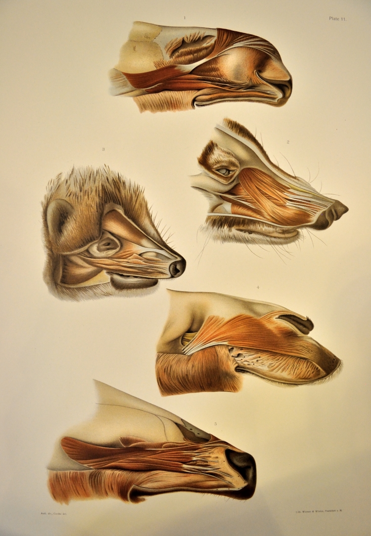
Comparative snoot muscle anatomy. From top to bottom: elk, coati, hedgehog, dromedary, and wapiti. (I admit to being a bit confused about the nomenclature here, as my understanding is that the elk and wapiti generally refer to the same animal — any ideas?) Image credit: Beinecke Rare Book & Manuscript Library / Curious Sengi.

Sometimes, it really isn’t your fault. You start off looking fabulous, but then time takes its inexorable toll. Rust never sleeps and you seriously need a nose job. Pangolin (Manis spp.) Image credit: Yale Peabody Museum / Curious Sengi.

Other times, the best intentions of your preparator just goes a bit awry. . . . Virginia opossum (Didelphis virginiana). Image credit: Yale Peabody Museum / Curious Sengi.

Then there are instances of– HOLY CRAP WHAT HAPPENED HERE?!? Malayan or Sunda Colugo (Cynocephalus variegatus). Image credit: Yale Peabody Museum / Curious Sengi.

This mole (Talpa spp.) is delighted you are here! Image credit: Cani Animali e Natura.
In celebration of Mole Day, we honor the life and research of Dr. Gillian Godfrey, who is largely remembered for her work on the life history of moles (the furry, digging kind) and writing popular books on the subject.
Gillian was described as “an extremely shy but fiercely dedicated zoologist”, who was drawn to ecology despite the “abominable lectures” given at Oxford University by Professor Charles S. Elton (1900 – 1991). After the term ended, she contacted Elton about joining his Bureau of Animal Population, but she did so with little expectation that she could have any hand in the scientific work going on there:
Her interview with Elton was awkward. He told her he didn’t care to have women in the Bureau just yet. She offered to work as a bottle washer and that did the trick. There wasn’t much future in bottle washing he retorted, so she had better come and do research (Crowcroft 1991).
Attitudes towards women in science in the 1950s was, at best, greeted with amusement or skepticism, but Gillian joined a group investigating vole (Microtus spp.) biology and pursued an ambitious research project on the “factors affecting the survival, movements, and intraspecific relations during early life” in vole populations (do not worry, the moles will come later).

Vole (Microtus spp.) Image credit: David Chapman via Saga Magazine.
The first step in this project was to induce the voles to nest so she could reliably return to her study subjects and trace their life histories.
With great single-mindedness, and almost no experience with hand tools, she set about mass producing nest boxes in D.K.’s [technician Denys Kempson’s] workshop. After his initial consternation, not because of the possibility of her injuring herself, but because he feared she might damage his tools, he diplomatically suggested that she work undisturbed in the field store. For many days the corridor echoed with the sounds of saw and hammer, and Gillian emerged with large numbers of wooden nest boxes with removable lids (Crowcroft 1991).
For all the work and enthusiasm poured into building nest boxes of all kinds of design, the voles were unappreciative of the effort and continued to built their own nests in clandestine locations (Crowcroft 1991). So Gillian proceeded to comb every square inch of her study site, crawling about on her hands and knees, parting the long tussock grass, finding plenty of old nests, and learning more about the private life of voles than this frustrating exercise initially promised (Crowcroft 1991; Chitty 1996). But this was no way to gather data for her project.
Then, in an inspired change of tactics brought about by the failure of the voles to use her nest boxes, she set out to trace their movements and find their nests by putting radioactive rings on their legs and finding them with a Geiger-Müller counter. She was greatly assisted on the technical side by a physicist with amorous ambitions which were fruitless and ill-conceived. Cobalt 60 wire was obtained. . . . before the Boss got wind of the project. He was pretty upset by her initiative, but saw that the technique had such great possibilities that she got away with it. This was the first time small mammals had been tracked in this fashion. . . . I [Crowcroft] still have some mental discomfort when I recall cutting up the wire for Gillian with two pairs of pliers, and rescuing bits that flew off by using the screaming Geiger counter (Crowcroft 1991).
The portable Geiger-counter-on-a-stick used to trace the location of voles through the thick grass. Image credit: Godfrey 1954.
It was a brilliant innovation, one Gillian claimed was inspired by the use of radioactive materials to track the movements of click beetles (Agriotes spp.) (Godfrey 1954). By mounting a Geiger counter on the end of a pole, it was possible to sweep large areas of habitat and locate an individual animal (Mellanby 1971). In writing up her novel methodology, Gillian acknowledged the advantage of this relatively non-invasive technique, since: “Nearly all available information about small wild animals has been obtained by indirect methods. . . .and it is usually impossible to assess the errors introduced. Trapping is frequently used in studies on movements but probably affects normal behaviour (Godfrey 1954).”
There were other breakthroughs to be had. Crowcroft (1991) writes:
I can recall finding Gillian in the vole room, hands streaming with blood and face streaked with tears, bravely pressing on with vole examinations, and explaining with great embarrassment, “Oh, but they bite so hard!” A few weeks later she was deftly holding them by the loose skin of the back with one hand, palpating the abdomen with the other.
One imagines that this determination and persistence carried Gillian through the tough years doing her doctorate. While composing her thesis, she was caught in the cross-currents of opposing views held by her examiners. Forced to write and re-write sections to appease these men who considered each other heretics, Gillian still navigated the conflicting torrents, and emerged triumphant. She was granted a doctorate by Oxford University in 1953 and was the first woman to complete such a degree in the Bureau of Animal Population (Crowcroft 1991; Chitty 1996).

What the radioactive tracking data look like for a single vole. For this given research project, Gillian collected 709 recordings of 23 animals. These maps showed the average distance between farthest points of capture was 29.04 yards (26.6 m), encompassing an area of 235.17 square yards (215.04 square meters). Data like these provide important information on the life history and distribution of these small mammals. Image credit: Godfrey 1954.

Moles are incredibly adapted to life digging underground: reduced, almost useless eyes, ears lacking any external pinnae, and powerful digging forelimbs. Since moles spend nearly all their lives underground, it had been almost impossible to reliably track their daily movements. Image credit: Mellanby 1971.
Gillian applied the same radioactive tagging technique to study the movement of moles (Talpa spp.), which are true insectivorans and not rodents like voles. This method was ideal for tracking these secretive animals. Instead of ringing the leg, a metal band with a soldered capsule containing radioactive Cobalt 60 was fixed to the mole’s conveniently club-shaped tail, which did not permit the ring to slip off when secured at the base. The ring could be detected up to 30 cm (1 ft) underground using the Geiger counter.

This drawing demonstrates the mole’s club-shaped tail: constricted at the base and widening towards the end. The black rectangle represents the position of the radioactive tracking ring. The shape of the tail prevented the ring from slipping off. Image credit: Mellanby 1971.
Even though Gillian’s tracking method became more sophisticated over the years, it was still a hazardous business to work with radioactive materials. Though she only tracked one animal at a time, she still had to be careful that the animals did not escape from the study site and leave radioactive rings strewn all over the English countryside. Of course, there was concern that prolonged exposure to Cobalt 60 would have a deleterious effect on the moles. Despite all these dangers, Gillian discovered a lot about these animals, including their reproductive habits, home range, and propensity towards three bouts of periodic activity over a 24 hour period (Mellanby 1971). In 1960, Gillian and her husband, Peter Crowcroft, a Tasmanian zoologist and zoo director (Chitty 1996), published The Life of the Mole, which received both academic and popular praise (Kettlewell 1961).

Image credit: Oxfam.

Image credit: Amazon.co.uk.
Unfortunately, Gillian Godfrey’s own trail quickly goes cold. Like many women of her time, Gillian was largely defined by her husband. She married Peter Crowcroft in 1952, while they were still students together at Oxford (Lidicker & Pucek 1997). We learn from an article in the Chicago Tribune that Peter was hired to be director of the Brookfield Zoo in 1968. At the time, Gillian was researching marsupials at the University of Adelaide, but would soon leave to join Peter in Chicago. Upon Peter’s death in 1996 at the age of 73, we read in his obituary that Gillian was, in fact, his second wife. But at the time of publication, she had disappeared from the picture and Peter was survived by another wife, Lisette.

Gillian seems to have retained her maiden name throughout her scientific career, but sometimes it was necessary to remind people to whom she was married. Image credit: Godfrey 1954.
What happened to Gillian? The answers are harder to find. Radioactive tagging is no longer used to track animal movements in the field, so it is quite likely this contributed to her work fading from scientific consciousness. But it was an ingenious solution to the problem of following shy, elusive animals in way that was least disruptive to their habits. Were there more ingenious solutions to address new questions that sparked her interest? I wish that I were able to find more information about Gillian’s later life and career, and that I could provide some kind of conclusion to this story.
So, Gillian, if you are out there, I hope you know that we think you are amazing!

Gillian out the field, listening for radioactive small mammals. Image credit: Godfrey 1954.
“Australian New Zoo Head at Brookfield.” Chicago Tribune. 7 February 1968: 4. Chicago Tribune. Web. Accessed 20 October 2016.
Chitty, Dennis. 1996. “Do Lemmings Commit Suicide? Beautiful Hypotheses and Ugly Facts.” New York, NY: Oxford University Press.
Crowcroft, Peter. 1991. Elton’s Ecologists: A History of the Bureau of Animal Population. Chicago, IL: The University of Chicago Press.
Kettlewell, H.B.D. 1961. “All about the mole.” New Scientist 9 (217): 107.
Lidicker, W.Z. & Z. Pucek. 1997. “William Peter Crowcroft (1922 – 1996).” Acta Theriologica 42 (3): 343 – 349.
Mellanby, Kenneth. 1971. The Mole. New York, NY: Taplinger Publishing Company.

Mondays are quiet here in the public galleries since the museum is closed to visitors. So we took the opportunity to squeeze through some tight wall spaces and clamber into an exhibit case for an official photo shoot for YPM VP 12048, the type specimen for Brontops robustus. The pebble-strewn ground is a lie. It is actually a perilous game of walking on thin ice: keep your weight on the bouncing planks of plywood, avoid falling through the chicken wire! Of course, all those darned loose rocks covering everything make it impossible to see the difference. . . . . . Image credit: Yale Peabody Museum / Curious Sengi.

Up close and personal. This animal belongs to the Family Brontotheriidae. Brontotheres are odd-toed ungulates (perissodactyls) and greatly resemble rhinos; however, modern analysis shows that this extinct lineage of mammals are most closely related to horses (Benton 2005). Image credit: Yale Peabody Museum / Curious Sengi.

Though a few areas of this skeleton may be reconstructed, what you see on display here is the real fossil! This well-preserved and largely complete specimen was discovered in 1875 near Chadron in the northwest corner of Nebraska in Oligocene aged deposits (Osborn 1929). Image credit: Yale Peabody Museum / Curious Sengi.
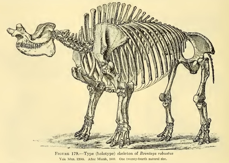
Brontotheres (sometimes called by their older name, titanotheres) are the subject of a richly illustrated, two-volume masterwork by American Museum paleontologist Henry Fairfield Osborn (1857 – 1935). The exact specimen on display today is reconstructed here in an illustration originally published by O.C. Marsh in 1889. Image credit: Osborn 1929.

The skeleton was actually mounted for display in 1916 under the direction of Richard Swann Lull (1867 – 1957), who studied under Osborn. Unlike the static standing pose in the Marsh illustration, Lull opted to capture the moment of a charging run, with the pert little tail in the air with rhino-like indignation. This exact same pose can be seen on display. When Lull measured the skeleton after it was mounted, he reported the height at the shoulder to be 8 feet, 2 1/2 inches (2.5 meters). Image credit: Osborn 1929.

Fanciful reconstruction of life in a brontothere herd. There used to be very different grazers out there on the Great Plains! Image credit: Restoration by Erwin Christman & Charles R. Knight, reproduced in Osborn 1929.
Benton, Michael. 2005. Vertebrate Paleontology, third edition. Oxford: Blackwell Publishing.
Osborn, Henry Fairfield. 1929. Titanotheres of Ancient Wyoming, Dakota, and Nebraska, Vol. 1. Washington, D.C.: United States Government Printing Of


#/media/File:La_Vierge_au_lys.jpg)